It is often very hard for non-operators to understand that the mixed liquor represents a mass of living organisms! This is the reason that so many people react as they do when viewing mixed liquor under a light microscope for the first time. It is not at all unusual for people to respond with surprise and excitement upon seeing activated sludge “bugs” (living organisms). Beyond the novelty of the light microscope lies one of the most powerful tools for assessing the activated sludge process that is available to operators. The ability to directly view the type and activity of the microorganisms involved in the process offers unparalleled insight into what is happening. As a further endorsement, the use of the light microscope is actually quite simple.
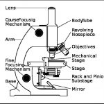
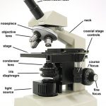
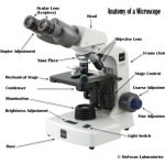
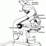
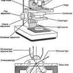
A modern light microscope is an instrument consisting of a light source, a stage where the object to be viewed is placed, an objective lens where light from the object is first magnified and an eyepiece, where the magnified image is again enlarged and viewed. Some provision for focusing the image, moving the viewed object around and controlling the light source is generally also provided. For activated sludge work, a microscope with objective lenses that will magnify to 4, 10, 40 and 100 power (X) are generally recommended. Most eyepieces provide an additional magnification of 10X, resulting in overall magnifications of 40, 100, 400 and 1000 times the original size of the object being viewed.
Samples of mixed liquor are best obtained from the settled sludge in a fresh settleometer test, or from a sample taken directly from the exit point of the aeration basin. For viewing live organisms, the sample should be as fresh as
Figure 10.19 – Schematic of a Compound Light Microscope with Built-it Light Source
possible. To prepare a sample to be viewed with a microscope, place a drop of the sample on a clean glass slide and cover it with a small, thin piece of glass, known as a cover slip. The cover slip prevents the sample from dying out too fast and prevents the lenses from accidentally contacting the wet sample. The light source should be adjusted in order to obtain the maximum contrast between light and dark. Too much light washes out the object, too little does not allow enough contrast to see details. Slides should be viewed first using the low power 4X objective lens.
Once an organism of interest is located, the higher power objectives can be used to discern greater detail. Be aware that the highest power objective lens, (100X), is used in conjunction with optical oil that is placed between the lens and the cover slip in order to allow a full field of view. (This practice is known as “oil immersion”). This 1000X magnification is generally only used when attempting to identify specific aspects of microorganisms, such as cell separations (septa) in filamentous bacteria.

